
The skin is an ecosystem. Isolated cell testing (monocultures) often fails to capture how active ingredients truly perform because cells rely on constant chemical communication (paracrine signaling) with their neighbors. To ensure complete physiological relevance, we utilize specialized co-culture models to mimic the true cellular environment of human skin.
1. Melanocyte & Keratinocyte Co-Culture (The Pigmentation Model)
Primary Cosmetic Claims: Advanced skin brightening, dark spot reduction, and hyperpigmentation control.
Testing melanin production in isolated melanocytes only tells half the story. In real human skin, a single melanocyte produces pigment (melanin) and physically transfers it to approximately 36 surrounding epidermal keratinocytes through arm-like structures called dendrites.
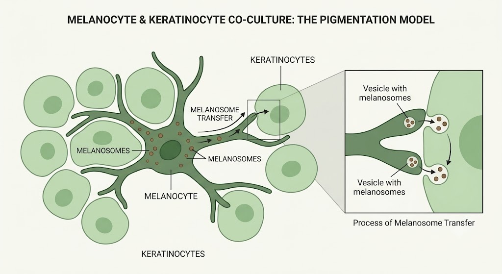
- Mechanism Tested: By co-culturing these two cell types together, we can evaluate an active ingredient’s ability to not only inhibit melanin synthesis but also block the physical transfer of melanosomes into the visible upper layers of the skin (often by antagonizing PAR-2 receptors).
- Value: Crucial for proving the efficacy of comprehensive brightening complexes that target multiple pathways in the melanogenesis cascade.
2. Fibroblast & Keratinocyte Co-Culture (The Dermal-Epidermal Junction)
Primary Cosmetic Claims: Deep wrinkle reduction, skin redensifying, wound healing, and structural barrier repair.
The Dermal-Epidermal Junction (DEJ) is the undulating membrane that connects the epidermis to the dermis below it. As we age, this junction flattens, reducing nutrient exchange and causing sagging. Fibroblasts (dermal cells) and keratinocytes (epidermal cells) must communicate constantly via growth factors (like TGF-β and KGF) to maintain this structure.
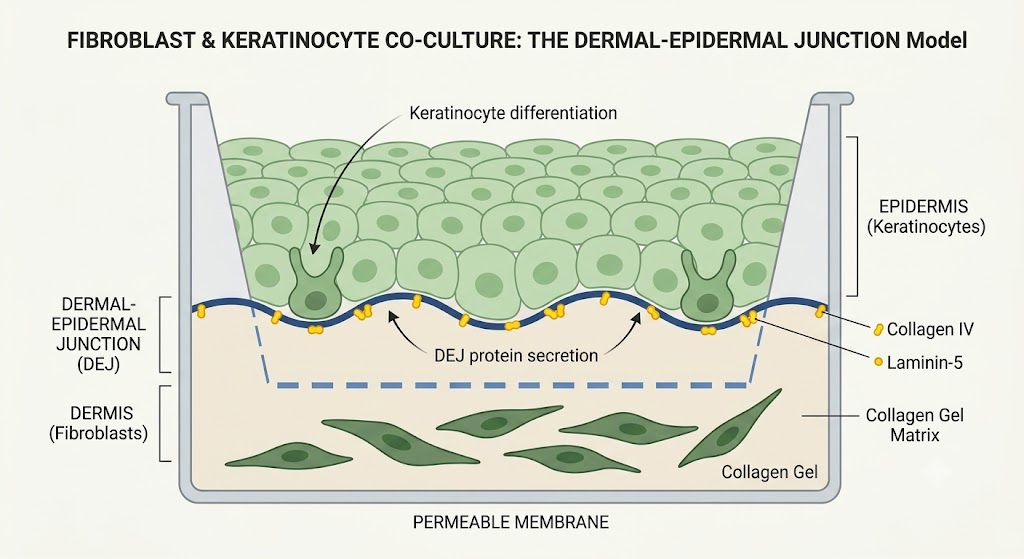
- Mechanism Tested: We culture epidermal cells above dermal cells to observe their biochemical cross-talk. We measure how an active applied to the “epidermis” signals the “dermis” to increase the synthesis of DEJ-specific anchoring proteins, such as Collagen IV, Collagen VII, and Laminin-5.
- Value: Provides robust substantiation for “pro-aging” and firming products by proving the formula successfully penetrates and stimulates the deep dermal layers through natural cellular communication.
3. Adipocyte & Fibroblast Co-Culture (The Hypodermis Model)
Primary Cosmetic Claims: Body contouring, anti-cellulite, skin firming, and targeted facial plumping (lipofilling).
The deepest layer of the skin (the hypodermis) is composed largely of adipocytes (fat cells). These cells don’t just store fat; they interact continuously with dermal fibroblasts to regulate skin tension, elasticity, and the extracellular matrix.
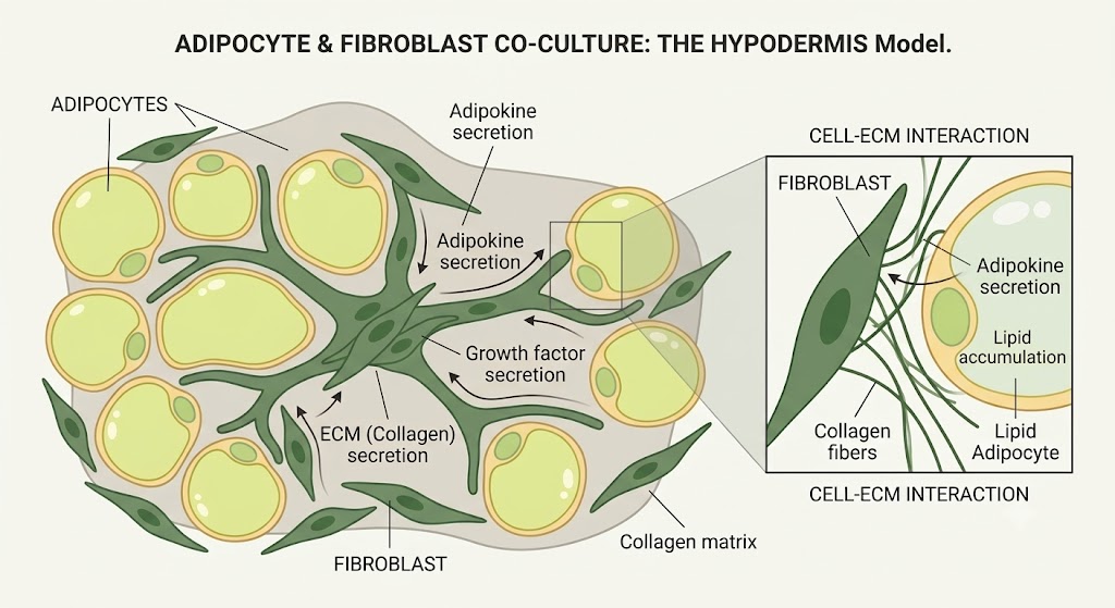
- Mechanism Tested: We co-culture human pre-adipocytes with fibroblasts to evaluate metabolic interactions. Depending on the brief, we can test for lipolysis (fat breakdown for slimming/anti-cellulite claims) or lipogenesis/adipogenesis (fat cell volume increase for facial plumping and wrinkle-filling claims).
- Value: Essential for body care formulations and advanced facial volumizers, proving that the active ingredients can successfully influence deep-tissue lipid metabolism and structural remodeling.
4. Nerve and Skin Tissue Interaction (Neuro-Cosmetics)
Primary Cosmetic Claims: Sensitive skin relief, anti-stinging, itch reduction, and “neuro-soothing.”
This is a highly advanced assay designed for the emerging field of neuro-cosmetics. Because skin is heavily innervated, cosmetic discomfort (stinging, burning) often stems from nerve-cell communication. We use sophisticated 2D or 3D co-culture models that integrate sensory neurons with keratinocytes or fibroblasts to observe their cross-talk.
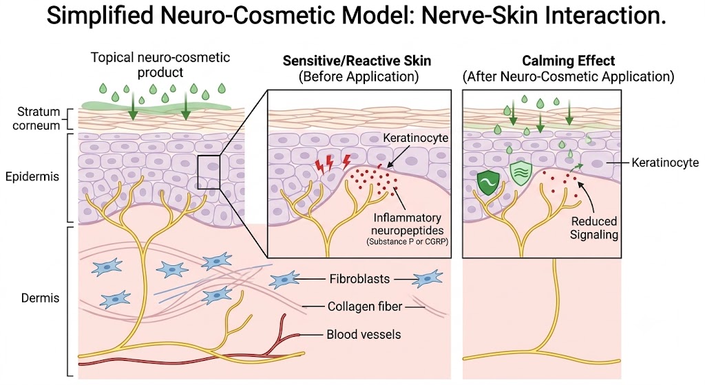
- Value: Essential for substantiating claims on products formulated for hyper-reactive, reactive, or sensitive skin types by proving the product successfully calms the skin’s neural network.
- By using these innervated models, we can substantiate high-value cosmetic claims by quantifying three critical mechanisms:
- Neuropeptide Release & Regulation (The “Soothing” Claim): When skin is stressed (e.g., by UV, chemicals, or temperature changes), nerves release pro-inflammatory neuropeptides like Substance P and CGRP, causing the sensations of stinging, itching, or burning. We measure a cosmetic active’s ability to intercept this communication and down-regulate these markers.
- Neurite Outgrowth (The “Neuro-Aging” Claim): As skin ages, the density of nerve endings decreases, leading to slower epidermal turnover and impaired wound healing. We use advanced imaging to measure the length and branching density of neurites (nerve fibers). Ingredients that stimulate neurite outgrowth (often via Nerve Growth Factor – NGF) support structural anti-aging and skin vitality.
5. Immune-Skin Co-Culture Models (The Sensitization Model)
The skin is our first line of immunological defense. When an ingredient triggers an allergic reaction or inflammation, it isn’t just the epidermal cells reacting—it is the skin’s resident immune cells sounding the alarm. To accurately test for true hypoallergenic and soothing properties, we must evaluate how cosmetic actives interact with the skin’s immune system.
We utilize advanced co-cultures combining human keratinocytes (epidermal cells) with macrophages (the sentinel immune cells of the skin tissue) to replicate this complex immunological cross-talk in vitro.
1. Substantiating “Hypoallergenic” & “Non-Sensitizing” Claims
The Challenge: Traditional “hypoallergenic” claims often rely solely on human patch testing (HRIPT), which occurs late in product development and only tells you if a reaction happened, not why.
How the Model Works:
We expose the macrophage-keratinocyte co-culture to the new cosmetic active or preservative system. If an ingredient is a potential sensitizer (allergen), it will trigger the keratinocytes to release danger signals, which in turn activate the macrophages into an aggressive, inflammatory state (M1 polarization).
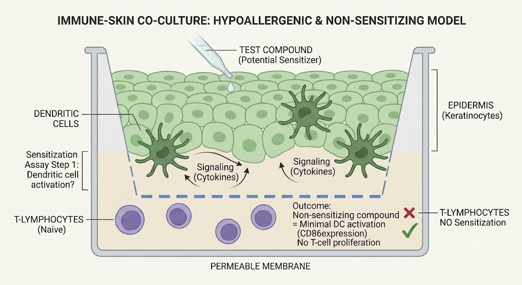
- Mechanism Tested: We quantify the release of specific sensitization biomarkers and pro-inflammatory cytokines (such as IL-18, IL-1α, and CD86 expression).
- Value: A lack of immune activation in this model provides robust, early-stage, cruelty-free proof that an ingredient is bio-compatible and truly hypoallergenic, saving brands from costly late-stage clinical failures.
2. Substantiating “Anti-Inflammatory” & “Soothing” Claims
The Challenge: Stressed skin (from UV rays, pollution, or harsh peels) creates a vicious cycle of inflammation. Brands need to prove their botanical extracts or peptides can actually break this cycle.
How the Model Works:
We use a “trans-well” indirect co-culture system where keratinocytes and macrophages share the same fluid environment but are separated by a porous membrane. We artificially induce stress in the skin cells (e.g., using chemical irritants or simulated UV radiation). The stressed skin cells send distress signals to the macrophages, triggering a massive inflammatory cascade. We then apply the soothing cosmetic active.
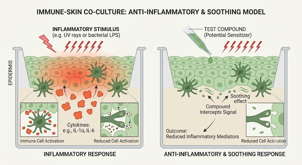
- Mechanism Tested: We measure the active ingredient’s ability to interrupt the biochemical cross-talk between the cells, specifically tracking the down-regulation of major inflammatory mediators like TNF-α, IL-6, and Prostaglandin E2 (PGE2). We also monitor the macrophages shifting from a pro-inflammatory (M1) state to a tissue-repairing (M2) state.
- Value: Delivers irrefutable, mechanism-of-action data for claims like “visibly reduces redness,” “calms reactive skin,” and “accelerates post-procedure recovery.”